Contents Yves Clermont, Michael Lalli, Zsuzsanna Bencsath-Makkai
LIGHT-MICROSCOPIC HISTOLOGY ATLAS
     2. Connective Tissue
  1     2     3     4     5     6     7     8     9     10     11     12     13     14     15     16     17     18     19  

    2.19   Multilocular adipocytes Version française
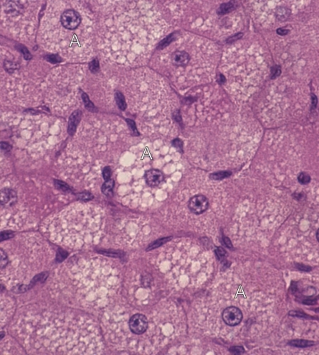
Section of the trachea of a dog.

This field shows a cluster of multilocular adipocytes (A). Some of these cells show their centrally located spherical nucleus. Their cytoplasm contains numerous lipid droplets of various sizes. This lipid was dissolved during the histological procedure.

The granularity of the cytoplasm between these vacuoles is due to the acidophilic mitochondria.

The elongated nuclei, seen between the adipocytes, belong to fibrocytes or endothelial cells of capillaries.

Stain: H–­­E
Magnification: ×900


We'd very much appreciate receiving your comments and suggestions!We'd very much appreciate receiving your comments and suggestions!

Creative Commons Licence The text and images of this Histology Atlas, by Yves Clermont, Michael Lalli & Zsuzsanna Bencsath-Makkai, are licensed under a Creative Commons Attribution-Noncommercial-No Derivative Works 2.5 Canada Licence and cannot be modified without the written permission of the authors. Use of any text or images must carry an acknowledgement which includes a link to the original work.


HAweb/Zs B-M -> HAbase-generated on 2013-09-13