Contents Yves Clermont, Michael Lalli, Zsuzsanna Bencsath-Makkai
LIGHT-MICROSCOPIC HISTOLOGY ATLAS
     2. Connective Tissue
  1     2     3     4     5     6     7     8     9     10     11     12     13     14     15     16     17     18     19  

    2.15   Reticular fibres Version française
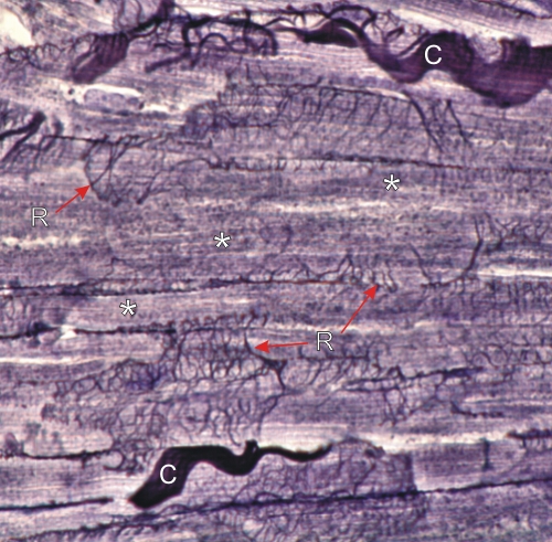
Section of a cardiac muscle stained with silver nitrate to show numerous reticular fibres in black.

These small fibres are composed of type III collagen rich in carbohydrates. These fine black reticular fibres, seen here in face view, form networks (R) that surround the individual bluish striated cardiac muscle fibres (*).

Between groups of muscle fibres there are also thick wavy type I collagen fibres (C) in a brownish-black colour.

Stain: Silver
Magnification: ×900


We'd very much appreciate receiving your comments and suggestions!We'd very much appreciate receiving your comments and suggestions!

Creative Commons Licence The text and images of this Histology Atlas, by Yves Clermont, Michael Lalli & Zsuzsanna Bencsath-Makkai, are licensed under a Creative Commons Attribution-Noncommercial-No Derivative Works 2.5 Canada Licence and cannot be modified without the written permission of the authors. Use of any text or images must carry an acknowledgement which includes a link to the original work.


HAweb/Zs B-M -> HAbase-generated on 2013-09-13