|
||
| 2. Connective Tissue | ||
| 1 2 3 4 5 6 7 8 9 10 11 12 13 14 15 16 17 18 19 |
| |||
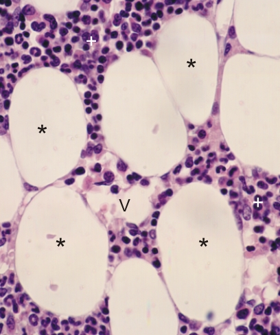 |
Section of the bone marrow.
This field shows, among the numerous blood cells in formation (+), several unilocular adipocytes (*). These cells have a large empty space occupied, in vivo, by a lipid droplet that was extracted with fat solvents. The thin peripheral cytoplasm of the adipocytes and the surrounding connective tissue fibrils are barely visible. A small vessel (V) is below the irregular space containing a few red blood cells. Stain: HE
|
||