Contents Yves Clermont, Michael Lalli, Zsuzsanna Bencsath-Makkai
LIGHT-MICROSCOPIC HISTOLOGY ATLAS
     2. Connective Tissue
  1     2     3     4     5     6     7     8     9     10     11     12     13     14     15     16     17     18     19  

    2.10   Mast cells in the connective tissue Version française
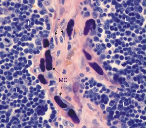
Section of a rat thymus stained with acid fuchsin and toluidine blue.

The nuclei and cytoplasm of lymphocytes (L) stain blue and mast cell (MC) granules stain purple with toluidine blue. These metachromatic granules are purple owing to the acidic glycoproteins present in heparin, an anticoagulant.

Several mast cells are seen in a connective tissue septum showing fibrocytes (F) and the acidophilic collagen.

Stain: Toluidine blue
Magnification: ×900


We'd very much appreciate receiving your comments and suggestions!We'd very much appreciate receiving your comments and suggestions!

Creative Commons Licence The text and images of this Histology Atlas, by Yves Clermont, Michael Lalli & Zsuzsanna Bencsath-Makkai, are licensed under a Creative Commons Attribution-Noncommercial-No Derivative Works 2.5 Canada Licence and cannot be modified without the written permission of the authors. Use of any text or images must carry an acknowledgement which includes a link to the original work.


HAweb/Zs B-M -> HAbase-generated on 2013-09-13