Contents Yves Clermont, Michael Lalli, Zsuzsanna Bencsath-Makkai
LIGHT-MICROSCOPIC HISTOLOGY ATLAS
     2. Connective Tissue
  1     2     3     4     5     6     7     8     9     10     11     12     13     14     15     16     17     18     19  

    2.12   Macrophages in connective tissue Version française
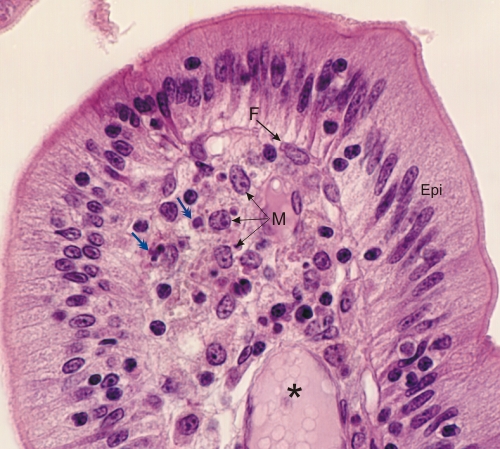
Section of an intestinal villus.

Underlying the simple columnar epithelium (Epi), the loose connective tissue shows several macrophages (M). These cells have a large nucleus in an abundant cytoplasm containing several basophilic or acidophilic inclusions of various sizes (double-headed arrows). These cellular residues have been phagocytized by the macrophages and are destined to be hydrolyzed by their lysosomes.

The nucleus of a fibrocyte (F) and a small blood vessel (*) are also labelled.

Stain: H–­­E
Magnification: ×900


We'd very much appreciate receiving your comments and suggestions!We'd very much appreciate receiving your comments and suggestions!

Creative Commons Licence The text and images of this Histology Atlas, by Yves Clermont, Michael Lalli & Zsuzsanna Bencsath-Makkai, are licensed under a Creative Commons Attribution-Noncommercial-No Derivative Works 2.5 Canada Licence and cannot be modified without the written permission of the authors. Use of any text or images must carry an acknowledgement which includes a link to the original work.


HAweb/Zs B-M -> HAbase-generated on 2013-09-13