Contents Yves Clermont, Michael Lalli, Zsuzsanna Bencsath-Makkai
LIGHT-MICROSCOPIC HISTOLOGY ATLAS
     2. Connective Tissue
  1     2     3     4     5     6     7     8     9     10     11     12     13     14     15     16     17     18     19  

    2.16   Adipose tissue Version française
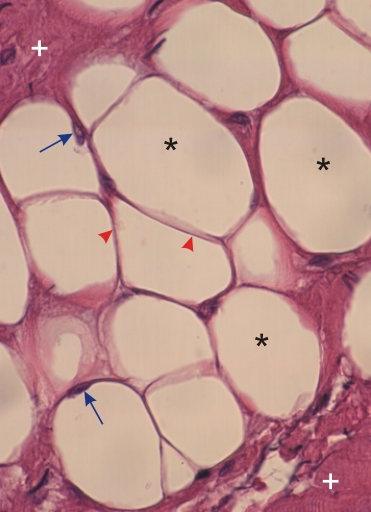
Section of adipose tissue from the wall of a larynx.

This field shows a group of unilocular adipose cells or adipocytes (*). These cells show a single large cavity normally occupied by a single large lipid droplet. The lipid was dissolved by the solvents utilized during the preparation of the histological section. The nuclei of these cells (arrows) are seen in a thin layer of cytoplasm at the periphery of the cells. Each adipocyte is surrounded by a thin layer of connective tissue (arrowheads).

Groups of adipocytes (*) are separated by large septae of connective tissue fibres (+).

Stain: H–­­E
Magnification: ×900


We'd very much appreciate receiving your comments and suggestions!We'd very much appreciate receiving your comments and suggestions!

Creative Commons Licence The text and images of this Histology Atlas, by Yves Clermont, Michael Lalli & Zsuzsanna Bencsath-Makkai, are licensed under a Creative Commons Attribution-Noncommercial-No Derivative Works 2.5 Canada Licence and cannot be modified without the written permission of the authors. Use of any text or images must carry an acknowledgement which includes a link to the original work.


HAweb/Zs B-M -> HAbase-generated on 2013-09-13