Sommaire Yves Clermont, Michael Lalli, Zsuzsanna Bencsath-Makkai
ATLAS D'HISTOLOGIE EN MICROSCOPIE OPTIQUE
     2. Tissus Conjonctifs
  1     2     3     4     5     6     7     8     9     10     11     12     13     14     15     16     17     18     19  

    2.7   Tissu conjonctif lâche English version
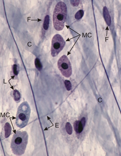
Mésentère du rat monté in toto et coloré au trichrome de Masson et à l’hématoxiline ferrique.

Ce champ de tissu conjonctif lâche montre, en haut et à droite, trois mastocytes (MC) dont le cytoplasme est chargé de granules autour d’un petit noyau sphérique. Les deux mastocytes en bas et à gauche sont dégranulés. Ces cellules sécrètent l’héparine un anticoagulant.

Sont également identifiés les éléments suivants: fibrocytes (F), leucocytes (L), fibres élastiques (E) et fibres de collagène (C).

Les noyaux des cellules mésothéliales ne sont pas inclus dans cette préparation.

Coloration: Trichrome de Masson
Grossissement: ×900


We'd very much appreciate receiving your comments and suggestions!We'd very much appreciate receiving your comments and suggestions!

Creative Commons Licence The text and images of this Histology Atlas, by Yves Clermont, Michael Lalli & Zsuzsanna Bencsath-Makkai, are licensed under a Creative Commons Attribution-Noncommercial-No Derivative Works 2.5 Canada Licence and cannot be modified without the written permission of the authors. Use of any text or images must carry an acknowledgement which includes a link to the original work.


HAweb/Zs B-M -> HAbase-generated on 2013-09-13