 |
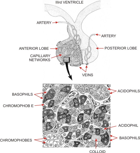
The posterior lobe contains cell processes of neurones, the cell bodies of which are located in the hypothalamic nuclei surrounding the third ventricle. These cells secrete a number of releasing factors that reach the capillaries or venules in the pituitary stalk which carry blood to the anterior lobe and stimulate the various endocrine cells present therein. Other secretory neurones send processes to the posterior lobe and release other hormones into the blood circulation. These include oxytocin, which acts on sensitized smooth muscle cells of the uterus and the myoepithelial cells of the mammary gland, and the antidiuretic hormone acting on the kidneys and controling water excretion.
In the anterior lobe (pars distalis), three main types of cells can be identified on the basis of their staining properties (bottom): - Acidophils, that secrete somatotrophin and others that secrete prolactin.
- Basophils, which include thyrotrophs secreting thyroid-stimulating hormone (TSH), corticotrophs secreting adrenocorticotrophin (ACTH), and gonadotrophs secreting either follicle-stimulating hormones (FSH, acting on the ovarian follicles) or luteinizing hormone (LH, involved in the formation and secretion of the corpus luteum or acting on the Leydig cells of the testis).
- Chromophobes, which are epithelial cells that do not stain with acidophilic or basophilic dyes. These cells are either reserve cells or degranulated acidophils and basophils.
Other cells, referred to as stellate or folliculo-stellate cells stain only lightly and have an undefined function. The three types of epithelial cells are not distributed at random throughout the anterior lobe, and clusters composed exclusively of acidophils (top right of bottom drawing) or of chromophobes (bottom left of bottom drawing) are regularly seen. Occasionally, the three types of cells are distributed around a small cavity containing a colloid, thus forming a cyst (bottom right of bottom drawing).
|