Contents Yves Clermont, Michael Lalli, Zsuzsanna Bencsath-Makkai
LIGHT-MICROSCOPIC HISTOLOGY ATLAS
     14. Endocrine Organs
  1     2     3     4     5     6     7     8     9     10     11     12     13     14     15     16     17     18     19     20     21     22     23     24     25  
  26     27     28     29     30  

    14.10   Thyroid follicles Version française
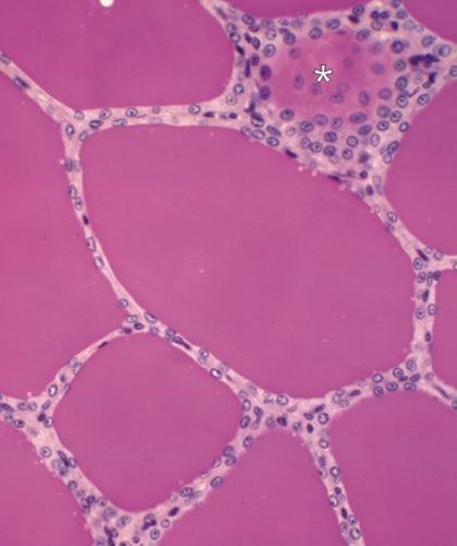
Thyroid follicles of a monkey.

In this case the epithelial cells are low cuboidal and the connective tissue between the follicles is barely visible. The well-fixed homogeneous colloid fills the lumen of the follicles.

The plane of section (top right) passes tangentially through the tip of a follicle (*) and some of the epithelial cells are seen side by side in a view from above.

Stain: H–­­E
Magnification: ×900


We'd very much appreciate receiving your comments and suggestions!We'd very much appreciate receiving your comments and suggestions!

Creative Commons Licence The text and images of this Histology Atlas, by Yves Clermont, Michael Lalli & Zsuzsanna Bencsath-Makkai, are licensed under a Creative Commons Attribution-Noncommercial-No Derivative Works 2.5 Canada Licence and cannot be modified without the written permission of the authors. Use of any text or images must carry an acknowledgement which includes a link to the original work.


HAweb/Zs B-M -> HAbase-generated on 2013-09-13