Contents Yves Clermont, Michael Lalli, Zsuzsanna Bencsath-Makkai
LIGHT-MICROSCOPIC HISTOLOGY ATLAS
     14. Endocrine Organs
  1     2     3     4     5     6     7     8     9     10     11     12     13     14     15     16     17     18     19     20     21     22     23     24     25  
  26     27     28     29     30  

    14.15   Human parathyroid Version française
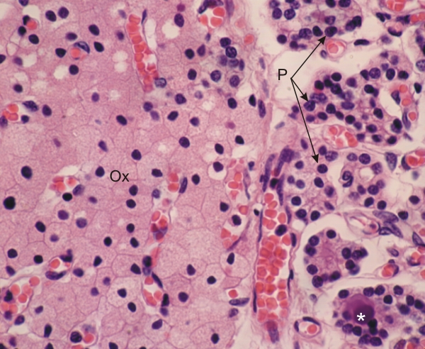
Section of a human parathyroid gland.

In this gland two main types of cells can be identified: the principal cells (P) and the oxyphils (Ox).

The principal cells are small and form irregular groups. They occasionally surround a small lumen, forming a cyst containing a colloid (*). The oxyphils have an abundant acidophilic cytoplasm due to the presence of numerous mitochondria.

Rare in younger individuals, the oxyphils increase in number with age.

In this section the numerous capillaries and venules containing red blood cells can be identified between groups of oxyphils and principal cells.

Stain: H–­­E
Magnification: ×1000


We'd very much appreciate receiving your comments and suggestions!We'd very much appreciate receiving your comments and suggestions!

Creative Commons Licence The text and images of this Histology Atlas, by Yves Clermont, Michael Lalli & Zsuzsanna Bencsath-Makkai, are licensed under a Creative Commons Attribution-Noncommercial-No Derivative Works 2.5 Canada Licence and cannot be modified without the written permission of the authors. Use of any text or images must carry an acknowledgement which includes a link to the original work.


HAweb/Zs B-M -> HAbase-generated on 2013-09-13