Contents Yves Clermont, Michael Lalli, Zsuzsanna Bencsath-Makkai
LIGHT-MICROSCOPIC HISTOLOGY ATLAS
     14. Endocrine Organs
  1     2     3     4     5     6     7     8     9     10     11     12     13     14     15     16     17     18     19     20     21     22     23     24     25  
  26     27     28     29     30  

    14.18   Adrenal cortex of a dog Version française
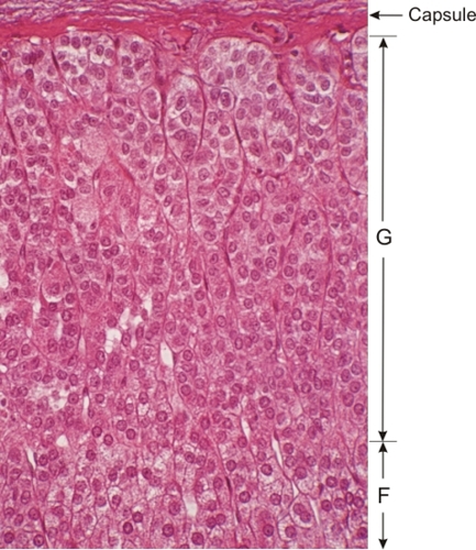
Section of a dog adrenal gland showing the superficial region of the adrenal cortex.

This field shows the zona glomerulosa (G) underlying the capsule and a small portion of the zona fasciculata (F) (bottom).

In the glomerulosa the glandular cells form irregular globular groups (suggested by the term glomerulosa). These groups of cells occasionally present an arched configuration. These cellular clusters are continuous with the cords or plates of the zona fasciculata. The boundary between these two zones is structurally imprecise.

In this preparation the collapsed vessels between the cords of glandular cells are barely visible, owing to the fixation procedure.

Stain: H–­­E
Magnification: ×600


We'd very much appreciate receiving your comments and suggestions!We'd very much appreciate receiving your comments and suggestions!

Creative Commons Licence The text and images of this Histology Atlas, by Yves Clermont, Michael Lalli & Zsuzsanna Bencsath-Makkai, are licensed under a Creative Commons Attribution-Noncommercial-No Derivative Works 2.5 Canada Licence and cannot be modified without the written permission of the authors. Use of any text or images must carry an acknowledgement which includes a link to the original work.


HAweb/Zs B-M -> HAbase-generated on 2013-09-13