|
||
| 14. Endocrine Organs | ||
| 1 2 3 4 5 6 7 8 9 10 11 12 13 14 15 16 17 18 19 20 21 22 23 24 25 | ||
| 26 27 28 29 30 |
| |||
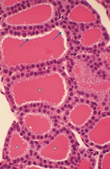 |
Thyroid gland of a dog.
This gland shows several sections of follicles. These follicles are lined with a simple cuboidal epithelium surrounding a cavity filled with an acidophilic colloid (*). The space between the apex of the glandular cells and the colloid (arrows) of some follicles is an artefact caused by the contraction of the colloid during the fixation of the organ. The follicles are separated by a small amount of connective tissue. Stain: HE
|
||