Contents Yves Clermont, Michael Lalli, Zsuzsanna Bencsath-Makkai
LIGHT-MICROSCOPIC HISTOLOGY ATLAS
     14. Endocrine Organs
  1     2     3     4     5     6     7     8     9     10     11     12     13     14     15     16     17     18     19     20     21     22     23     24     25  
  26     27     28     29     30  

    14.23   Pancreatic islet of Langerhans Version française
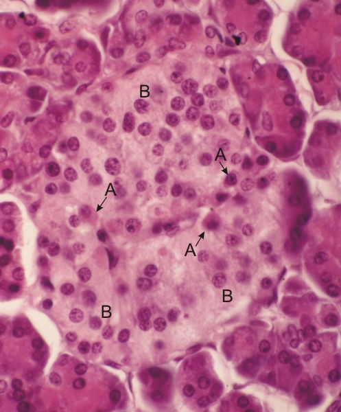
Section of a pancreatic islet of Langerhans from a monkey.

The exocrine acini of the pancreas are seen around the islet.

In this islet the closely packed glandular cells are generally lightly stained. Some of these cells are acidophilic and correspond to the acidophils (A) which secrete glucagon. The lightly stained cells correspond to the basophils (B) which secrete insulin.

The other cells of the islet, that is, the D cells, which secrete somatostatin, and the F cells, which secrete a specific pancreatic polypeptide, can be demonstrated only with immunocytochemical methods.

The capillaries associated with the islet are barely visible in this section.

Stain: H–­­E
Magnification: ×600


We'd very much appreciate receiving your comments and suggestions!We'd very much appreciate receiving your comments and suggestions!

Creative Commons Licence The text and images of this Histology Atlas, by Yves Clermont, Michael Lalli & Zsuzsanna Bencsath-Makkai, are licensed under a Creative Commons Attribution-Noncommercial-No Derivative Works 2.5 Canada Licence and cannot be modified without the written permission of the authors. Use of any text or images must carry an acknowledgement which includes a link to the original work.


HAweb/Zs B-M -> HAbase-generated on 2013-09-13