|
||
| 14. Glandes Endocrines | ||
| 1 2 3 4 5 6 7 8 9 10 11 12 13 14 15 16 17 18 19 20 21 22 23 24 25 | ||
| 26 27 28 29 30 |
| |||
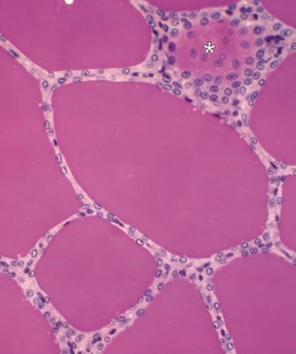 |
Follicules thyroïdiens dun singe. Dans ce cas les cellules folliculaires sont aplaties et le tissu conjonctif entre les follicules est à peine visible. La colloïde, bien fixée, est homogène et remplit complètement la lumière des follicules. (En haut et à gauche) Limage montre lextrémité dun follicule coupé tangentiellement (*). On y voit des cellules épithéliales côte-à-côte en vue plongeante. Coloration: HÉ
|
||