|
||
| 11. Cavité Orale | ||
| 1 2 3 4 5 6 7 8 9 10 11 12 13 14 15 16 17 18 19 20 21 22 23 24 25 | ||
| 26 27 28 29 30 31 32 33 34 35 36 37 38 39 40 41 |
| |||
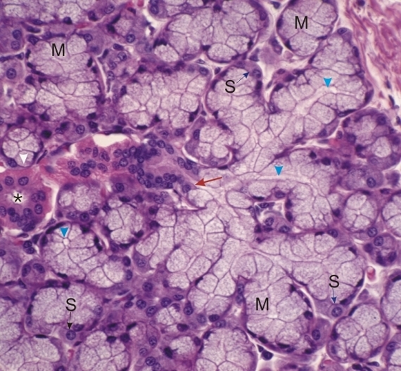 |
Glande sublinguale. Plusieurs acinus mixtes montrent une étroite lumière (pointes de flèches). Ces canalicules débouchent dans un canal intercalaire (flèche rouge). Les cellules muqueuses (M) des acinus mixtes peuvent être distinguées facilement des cellules séreuses (S). Un petit canal intralobulaire strié (*) est présent à proximité. Coloration: HÉ
|
||