Sommaire Yves Clermont, Michael Lalli, Zsuzsanna Bencsath-Makkai
ATLAS D'HISTOLOGIE EN MICROSCOPIE OPTIQUE
     11. Cavité Orale
  1     2     3     4     5     6     7     8     9     10     11     12     13     14     15     16     17     18     19     20     21     22     23     24     25  
  26     27     28     29     30     31     32     33     34     35     36     37     38     39     40     41  

    11.35   Cavité buccale: formation de la dent English version
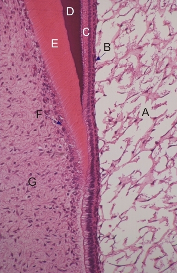
Coupe montrant la croissance de la couronne d’une incisive. Les structures suivantes son identifiées:
  1. Réticulum étoilé
  2. Couche intermédiaire
  3. Couche d’améloblastes
  4. Émail basophile en formation
  5. Dentine éosinophile en formation
  6. Odontoblastes
  7. Tissu conjonctif de la pulpe dentaire

Coloration: H–­­É
Grossissement: ×450


We'd very much appreciate receiving your comments and suggestions!We'd very much appreciate receiving your comments and suggestions!

Creative Commons Licence The text and images of this Histology Atlas, by Yves Clermont, Michael Lalli & Zsuzsanna Bencsath-Makkai, are licensed under a Creative Commons Attribution-Noncommercial-No Derivative Works 2.5 Canada Licence and cannot be modified without the written permission of the authors. Use of any text or images must carry an acknowledgement which includes a link to the original work.


HAweb/Zs B-M -> HAbase-generated on 2013-09-13