Sommaire Yves Clermont, Michael Lalli, Zsuzsanna Bencsath-Makkai
ATLAS D'HISTOLOGIE EN MICROSCOPIE OPTIQUE
     11. Cavité Orale
  1     2     3     4     5     6     7     8     9     10     11     12     13     14     15     16     17     18     19     20     21     22     23     24     25  
  26     27     28     29     30     31     32     33     34     35     36     37     38     39     40     41  
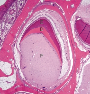
    11.32   Cavité buccale: bourgeon dentaire English version
Coupe d’un bourgeon dentaire d’un jeune singe.

Ce bourgeon est logé dans une cavité entouré d’os alvéolaire (+).

Il montre un stade initial de la formation de la couronne de la dent. Les éléments suivants sont identifiés:

  • L’organe de l’émail (*) qui coiffe le bourgeon
  • Couche basophile de l’émail (E)
  • Couche acidophile de dentine (D)

Ces trois couches entourent partiellement le tissu conjonctif de la pulpe ou papille (P) du bourgeon.

À ce stade la formation de la racine de la dent est à peine amorcée.

Coloration: H–­­É
Grossissement: ×25


We'd very much appreciate receiving your comments and suggestions!We'd very much appreciate receiving your comments and suggestions!

Creative Commons Licence The text and images of this Histology Atlas, by Yves Clermont, Michael Lalli & Zsuzsanna Bencsath-Makkai, are licensed under a Creative Commons Attribution-Noncommercial-No Derivative Works 2.5 Canada Licence and cannot be modified without the written permission of the authors. Use of any text or images must carry an acknowledgement which includes a link to the original work.


HAweb/Zs B-M -> HAbase-generated on 2013-09-13