|
||
| 11. Cavité Orale | ||
| 1 2 3 4 5 6 7 8 9 10 11 12 13 14 15 16 17 18 19 20 21 22 23 24 25 | ||
| 26 27 28 29 30 31 32 33 34 35 36 37 38 39 40 41 |
| |||
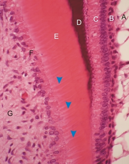 |
Incisive en formation dun singe. Les éléments suivants sont étiquetés:
Coloration: HÉ
|
||
|
||
| 11. Cavité Orale | ||
| 1 2 3 4 5 6 7 8 9 10 11 12 13 14 15 16 17 18 19 20 21 22 23 24 25 | ||
| 26 27 28 29 30 31 32 33 34 35 36 37 38 39 40 41 |
| |||
|
|
Incisive en formation dun singe. Les éléments suivants sont étiquetés:
Coloration: HÉ
|
||
![]() The text and images of this Histology Atlas, by Yves Clermont,
Michael Lalli & Zsuzsanna Bencsath-Makkai,
are licensed under a
Creative Commons Attribution-Noncommercial-No Derivative Works 2.5 Canada Licence
and cannot be modified without the written permission of the authors.
Use of any text or images must carry an acknowledgement which includes a link to the original work.
The text and images of this Histology Atlas, by Yves Clermont,
Michael Lalli & Zsuzsanna Bencsath-Makkai,
are licensed under a
Creative Commons Attribution-Noncommercial-No Derivative Works 2.5 Canada Licence
and cannot be modified without the written permission of the authors.
Use of any text or images must carry an acknowledgement which includes a link to the original work.