Sommaire Yves Clermont, Michael Lalli, Zsuzsanna Bencsath-Makkai
ATLAS D'HISTOLOGIE EN MICROSCOPIE OPTIQUE
     11. Cavité Orale
  1     2     3     4     5     6     7     8     9     10     11     12     13     14     15     16     17     18     19     20     21     22     23     24     25  
  26     27     28     29     30     31     32     33     34     35     36     37     38     39     40     41  
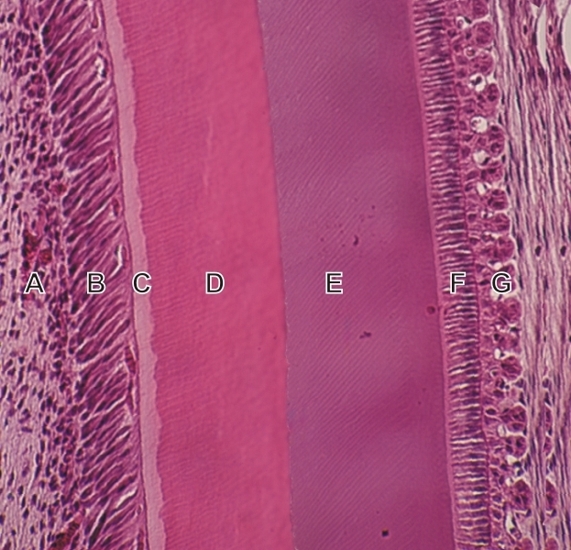
    11.40   Cavité buccale: formation de la dent English version
Incisive en croissance d’un rongeur.

Ce champ provient de la portion médiane de la longue racine en croissance continue de l’incisive.

Les couches ou structures suivantes sont identifiées (de gauche à droite):

  1. Pulpe dentaire
  2. Rangée d’odontoblastes allongés
  3. Prédentine non-minéralisée
  4. Dentine minéralisée
  5. Émail
  6. Rangée d’améloblastes sécrétoires
  7. Résidus de l’organe de l’émail

Coloration: H–­­É
Grossissement: ×300


We'd very much appreciate receiving your comments and suggestions!We'd very much appreciate receiving your comments and suggestions!

Creative Commons Licence The text and images of this Histology Atlas, by Yves Clermont, Michael Lalli & Zsuzsanna Bencsath-Makkai, are licensed under a Creative Commons Attribution-Noncommercial-No Derivative Works 2.5 Canada Licence and cannot be modified without the written permission of the authors. Use of any text or images must carry an acknowledgement which includes a link to the original work.


HAweb/Zs B-M -> HAbase-generated on 2013-09-13