Sommaire Yves Clermont, Michael Lalli, Zsuzsanna Bencsath-Makkai
ATLAS D'HISTOLOGIE EN MICROSCOPIE OPTIQUE
     11. Cavité Orale
  1     2     3     4     5     6     7     8     9     10     11     12     13     14     15     16     17     18     19     20     21     22     23     24     25  
  26     27     28     29     30     31     32     33     34     35     36     37     38     39     40     41  
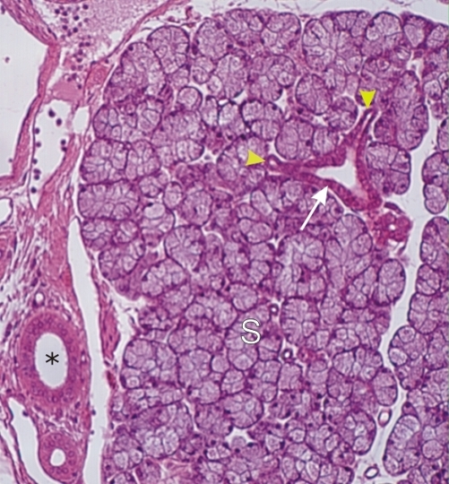
    11.13   Cavité buccale:glande salivaire English version
Coupe d’une parotide.

Une petite portion d’un lobule est à proximité d’une travée conjonctive. Cette travée contient un petit canal excréteur interlobulaire (*).

Parmi les acinus, composés exclusivement de cellules séreuses (S), on note la présence d’un canal intralobulaire (flèche). Ce canal est continu avec deux petits canaux intercalaires (pointes de flèches).

Coloration: H–­­É
Grossissement: ×300


We'd very much appreciate receiving your comments and suggestions!We'd very much appreciate receiving your comments and suggestions!

Creative Commons Licence The text and images of this Histology Atlas, by Yves Clermont, Michael Lalli & Zsuzsanna Bencsath-Makkai, are licensed under a Creative Commons Attribution-Noncommercial-No Derivative Works 2.5 Canada Licence and cannot be modified without the written permission of the authors. Use of any text or images must carry an acknowledgement which includes a link to the original work.


HAweb/Zs B-M -> HAbase-generated on 2013-09-13