Sommaire Yves Clermont, Michael Lalli, Zsuzsanna Bencsath-Makkai
ATLAS D'HISTOLOGIE EN MICROSCOPIE OPTIQUE
     11. Cavité Orale
  1     2     3     4     5     6     7     8     9     10     11     12     13     14     15     16     17     18     19     20     21     22     23     24     25  
  26     27     28     29     30     31     32     33     34     35     36     37     38     39     40     41  

    11.15   Oral cavity: salivary gland English version
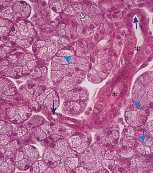
Glande parotide.

Les acinus montrent les cellules séreuses plus ou moins pyramidales autour d’une étroite lumière centrale (pointes de flèches).

Une coupe longitudinale d’un canal intralobulaire acidophile strié (*) est connecté à d’étroits canaux intercalaires (flèches).

Coloration: H–­­É
Grossissement: ×1000


We'd very much appreciate receiving your comments and suggestions!We'd very much appreciate receiving your comments and suggestions!

Creative Commons Licence The text and images of this Histology Atlas, by Yves Clermont, Michael Lalli & Zsuzsanna Bencsath-Makkai, are licensed under a Creative Commons Attribution-Noncommercial-No Derivative Works 2.5 Canada Licence and cannot be modified without the written permission of the authors. Use of any text or images must carry an acknowledgement which includes a link to the original work.


HAweb/Zs B-M -> HAbase-generated on 2013-09-13