Sommaire Yves Clermont, Michael Lalli, Zsuzsanna Bencsath-Makkai
ATLAS D'HISTOLOGIE EN MICROSCOPIE OPTIQUE
     11. Cavité Orale
  1     2     3     4     5     6     7     8     9     10     11     12     13     14     15     16     17     18     19     20     21     22     23     24     25  
  26     27     28     29     30     31     32     33     34     35     36     37     38     39     40     41  

    11.41   Cavité buccale: formation de la dent English version
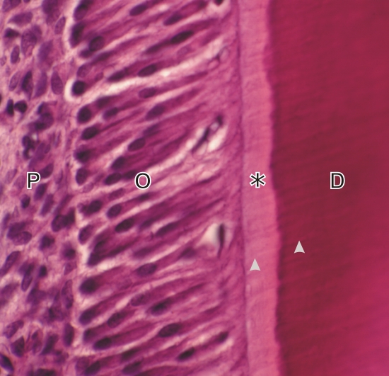
Incisive en croissance de rongeur.

Ce champ montre la pulpe dentaire (P) et à proximité une rangée d’odontoblastes (O) en contact avec la prédentine non-minéralisée (*). La dentine (D) minéralisée est très acidophile.

Des tubules de la dentine (pointes de flèches), qui contiennent les fins prolongements cytoplasmiques des odontoblastes, sont identifiables dans la prédentine et la dentine.

Coloration: H–­­É
Grossissement: ×1000


We'd very much appreciate receiving your comments and suggestions!We'd very much appreciate receiving your comments and suggestions!

Creative Commons Licence The text and images of this Histology Atlas, by Yves Clermont, Michael Lalli & Zsuzsanna Bencsath-Makkai, are licensed under a Creative Commons Attribution-Noncommercial-No Derivative Works 2.5 Canada Licence and cannot be modified without the written permission of the authors. Use of any text or images must carry an acknowledgement which includes a link to the original work.


HAweb/Zs B-M -> HAbase-generated on 2013-09-13