Sommaire Yves Clermont, Michael Lalli, Zsuzsanna Bencsath-Makkai
ATLAS D'HISTOLOGIE EN MICROSCOPIE OPTIQUE
     11. Cavité Orale
  1     2     3     4     5     6     7     8     9     10     11     12     13     14     15     16     17     18     19     20     21     22     23     24     25  
  26     27     28     29     30     31     32     33     34     35     36     37     38     39     40     41  
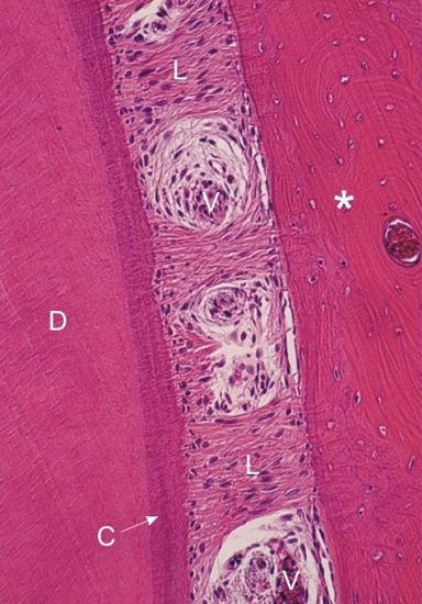
    11.28   Cavité buccale: ligament périodontal English version
Racine d’une dent.

Cette photo montre la dentine (D) et l’os (*) ainsi que le ligament périodontal (L) qui fait le pont entre les deux. La couche de cément acellulaire (C), légèrement basophile, recouvre la dentine de la racine.

Ce ligament periodontal est composé de fibres de collagène de type I et de nombreux fibrocytes. Les fibres conjonctives du ligament périodontal s’insèrent dans ce cément acellulaire et la matrice de l’os.

Le ligament périodontal montre des régions de tissu conjonctif lâche qui contiennent des petits vaisseaux (V).

Coloration: H–­­É
Grossissement: ×350


We'd very much appreciate receiving your comments and suggestions!We'd very much appreciate receiving your comments and suggestions!

Creative Commons Licence The text and images of this Histology Atlas, by Yves Clermont, Michael Lalli & Zsuzsanna Bencsath-Makkai, are licensed under a Creative Commons Attribution-Noncommercial-No Derivative Works 2.5 Canada Licence and cannot be modified without the written permission of the authors. Use of any text or images must carry an acknowledgement which includes a link to the original work.


HAweb/Zs B-M -> HAbase-generated on 2013-09-13