|
||
| 12. Appareil Digestif | ||
| 1 2 3 4 5 6 7 8 9 10 11 12 13 14 15 16 17 18 19 20 21 22 23 24 25 | ||
| 26 27 28 29 30 31 32 33 34 35 36 37 38 39 40 41 42 43 44 45 46 47 48 49 50 | ||
| 51 52 53 54 55 56 57 58 59 60 61 62 63 64 65 66 67 68 69 70 71 72 73 74 75 | ||
| 76 77 78 79 80 81 82 83 84 85 86 |
| |||
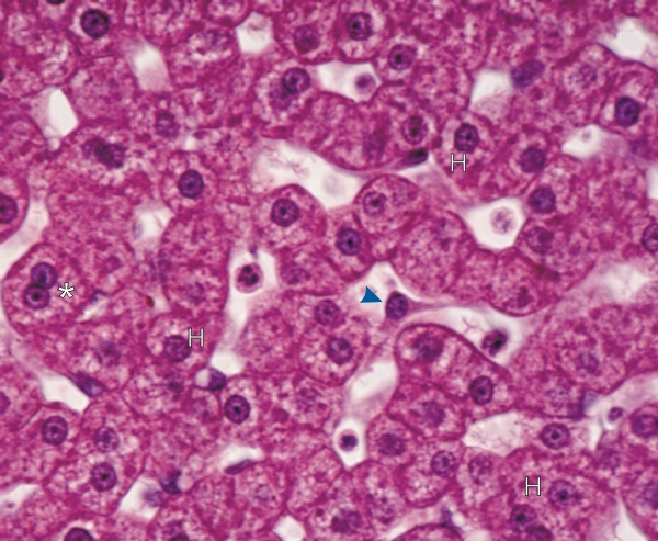 |
Coupe du foie dun singe. Les hépatocytes polyhédriques contiennent un noyau sphérique central. Certains hépatocytes sont binuclées (*). Les noyaux sont entourés dun cytoplasme abondant daspect granulaire. Ces granulations reflètent la multiplicité des organites cytoplasmiques (mitochondries, lysosomes, peroxisomes, glycogen, etc.). Les espaces sinusoïdaux entre les lames dhépatocytes sont delimités par des cellules endothéliales très minces et indistinctes et des cellules de Kupffer étoilées (pointe de flèche). Les petits canalicules biliaires sont également difficiles à identifier sur cette coupe épaisse. Coloration: HÉ
|
||