|
||
| 12. Appareil Digestif | ||
| 1 2 3 4 5 6 7 8 9 10 11 12 13 14 15 16 17 18 19 20 21 22 23 24 25 | ||
| 26 27 28 29 30 31 32 33 34 35 36 37 38 39 40 41 42 43 44 45 46 47 48 49 50 | ||
| 51 52 53 54 55 56 57 58 59 60 61 62 63 64 65 66 67 68 69 70 71 72 73 74 75 | ||
| 76 77 78 79 80 81 82 83 84 85 86 |
| |||
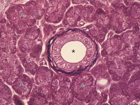 |
Coupe du pancréas dun chien. Un petit canal intralobulaire (*) montre un épithélium cubique simple encerclé dune mince couche conjonctive composée de fibres de collagènes acidophiles et de fibres élastiques colorées en noir (flèche blanche). Plusieurs acinus (A) entourent ce canal. Coloration: Hématoxyline Ferrique-É
|
||