Sommaire Yves Clermont, Michael Lalli, Zsuzsanna Bencsath-Makkai
ATLAS D'HISTOLOGIE EN MICROSCOPIE OPTIQUE
     12. Appareil Digestif
  1     2     3     4     5     6     7     8     9     10     11     12     13     14     15     16     17     18     19     20     21     22     23     24     25  
  26     27     28     29     30     31     32     33     34     35     36     37     38     39     40     41     42     43     44     45     46     47     48     49     50  
  51     52     53     54     55     56     57     58     59     60     61     62     63     64     65     66     67     68     69     70     71     72     73     74     75  
  76     77     78     79     80     81     82     83     84     85     86  
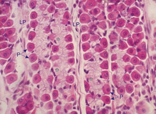
    12.14   Estomac English version
Muqueuse gastrique d’un chien.

Ce champ montre le collet des glandes gastriques et leurs mombreuses cellules pariétales (P) très acidophiles.

Ces cellules sont en présence des cellules muqueuses du collet (*) peu colorées et aux contours imprécis.

La lamina propria (LP) sépare les glandes.

Coloration: H–­­É
Grossissement: ×800


We'd very much appreciate receiving your comments and suggestions!We'd very much appreciate receiving your comments and suggestions!

Creative Commons Licence The text and images of this Histology Atlas, by Yves Clermont, Michael Lalli & Zsuzsanna Bencsath-Makkai, are licensed under a Creative Commons Attribution-Noncommercial-No Derivative Works 2.5 Canada Licence and cannot be modified without the written permission of the authors. Use of any text or images must carry an acknowledgement which includes a link to the original work.


HAweb/Zs B-M -> HAbase-generated on 2013-09-13