|
||
| 12. Appareil Digestif | ||
| 1 2 3 4 5 6 7 8 9 10 11 12 13 14 15 16 17 18 19 20 21 22 23 24 25 | ||
| 26 27 28 29 30 31 32 33 34 35 36 37 38 39 40 41 42 43 44 45 46 47 48 49 50 | ||
| 51 52 53 54 55 56 57 58 59 60 61 62 63 64 65 66 67 68 69 70 71 72 73 74 75 | ||
| 76 77 78 79 80 81 82 83 84 85 86 |
| |||
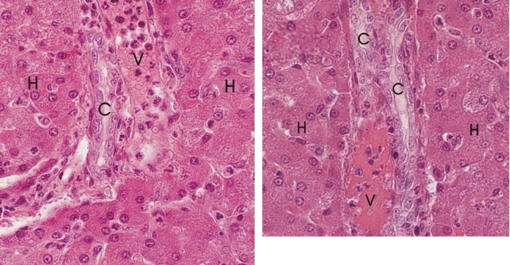 |
Ces deux champs du foie dun chien montrent des petits espaces portes entourés dhépatocytes (H). Chaque champ montre un cholangiole (C) dont la lumière est bordée de cellules épithéliales allongées et aplaties. À proximité, une petites veinules portes (V) contient des cellules sanguines. Coloration: HÉ
|
||