Sommaire Yves Clermont, Michael Lalli, Zsuzsanna Bencsath-Makkai
ATLAS D'HISTOLOGIE EN MICROSCOPIE OPTIQUE
     12. Appareil Digestif
  1     2     3     4     5     6     7     8     9     10     11     12     13     14     15     16     17     18     19     20     21     22     23     24     25  
  26     27     28     29     30     31     32     33     34     35     36     37     38     39     40     41     42     43     44     45     46     47     48     49     50  
  51     52     53     54     55     56     57     58     59     60     61     62     63     64     65     66     67     68     69     70     71     72     73     74     75  
  76     77     78     79     80     81     82     83     84     85     86  
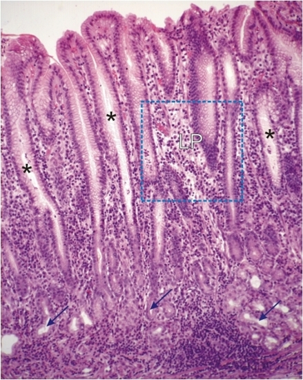
    12.25   Pylore English version
Coupe d’un pylore humain.

Dans cette muqueuse les cryptes (*) sont profondes alors que les glandes pyloriques tubulaires (flèches) sont courtes.

La lamina propria (LP) formée d’un abondant tissu conjonctif contient de nombreuses cellules lymphoïdes.

Voir le détail du territoire encadré dans la figure 12.26.

Coloration: H–­­É
Grossissement: ×265


We'd very much appreciate receiving your comments and suggestions!We'd very much appreciate receiving your comments and suggestions!

Creative Commons Licence The text and images of this Histology Atlas, by Yves Clermont, Michael Lalli & Zsuzsanna Bencsath-Makkai, are licensed under a Creative Commons Attribution-Noncommercial-No Derivative Works 2.5 Canada Licence and cannot be modified without the written permission of the authors. Use of any text or images must carry an acknowledgement which includes a link to the original work.


HAweb/Zs B-M -> HAbase-generated on 2013-09-13