|
||
| 12. Appareil Digestif | ||
| 1 2 3 4 5 6 7 8 9 10 11 12 13 14 15 16 17 18 19 20 21 22 23 24 25 | ||
| 26 27 28 29 30 31 32 33 34 35 36 37 38 39 40 41 42 43 44 45 46 47 48 49 50 | ||
| 51 52 53 54 55 56 57 58 59 60 61 62 63 64 65 66 67 68 69 70 71 72 73 74 75 | ||
| 76 77 78 79 80 81 82 83 84 85 86 |
| |||
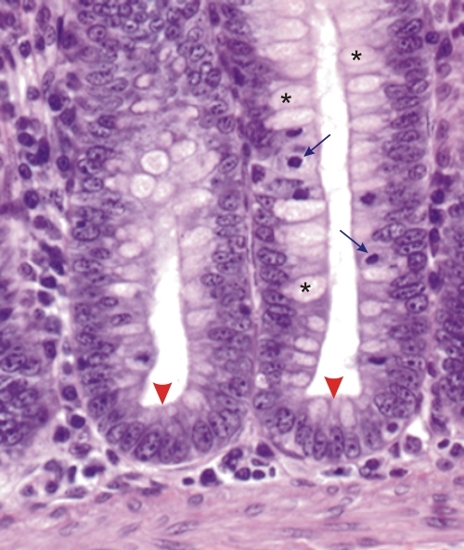 |
Muqueuses du côlon dun chien. Les bases de deux glandes intestinales montrent des cellules épithéliales indifférenciées (pointes de flèches) dont certaines sont en mitose (flèches). Parmi ces cellules certaines montrent les premiers signes de leur différenciation en cellules caliciformes (*). Coloration: HÉ
|
||