Contents Yves Clermont, Michael Lalli, Zsuzsanna Bencsath-Makkai
LIGHT-MICROSCOPIC HISTOLOGY ATLAS
     3. Muscle Tissue
  1     2     3     4     5     6     7     8     9     10     11     12     13     14     15     16     17  

    3.8   Striated muscle Version française
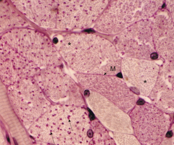
Cross section of striated muscle stained with periodic acid-Schiff (PAS) and hematoxylin.

This field shows the irregular polyhedral profiles of the muscle fibres with lightly stained transverse sections of myofibrils (*). Interspersed in the cytoplasm of some cells, the purple granulations correspond to accumulations of glycogen. Note the variations in the numbers of purple glycogen particles (arrows) in the cytoplasm of the various muscle cells.

The PAS technique also stains the basement membrane (M), with its abundant carbohydrate-rich type III collagen. This thin amorphous layer is closely associated with the cytoplasmic membrane (not visible) of muscle cells.

Stain: PAS-Hematoxylin
Magnification: ×900


We'd very much appreciate receiving your comments and suggestions!We'd very much appreciate receiving your comments and suggestions!

Creative Commons Licence The text and images of this Histology Atlas, by Yves Clermont, Michael Lalli & Zsuzsanna Bencsath-Makkai, are licensed under a Creative Commons Attribution-Noncommercial-No Derivative Works 2.5 Canada Licence and cannot be modified without the written permission of the authors. Use of any text or images must carry an acknowledgement which includes a link to the original work.


HAweb/Zs B-M -> HAbase-generated on 2013-09-13