Contents Yves Clermont, Michael Lalli, Zsuzsanna Bencsath-Makkai
LIGHT-MICROSCOPIC HISTOLOGY ATLAS
     3. Muscle Tissue
  1     2     3     4     5     6     7     8     9     10     11     12     13     14     15     16     17  

    3.5   Striated muscle Version française
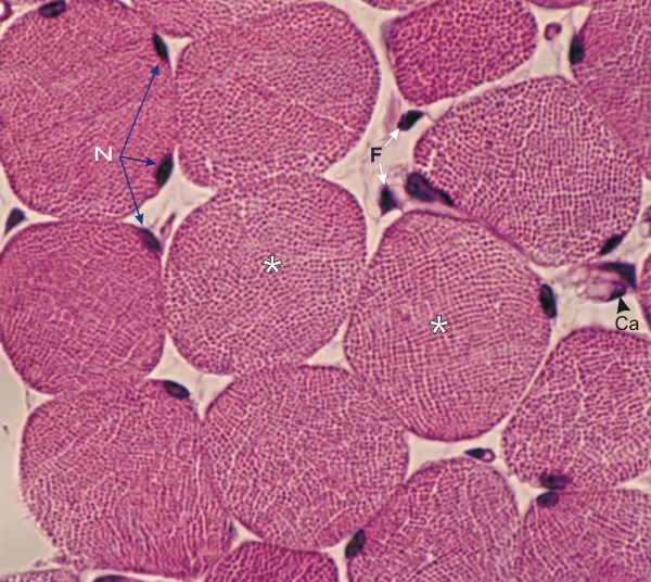
Cross sections of striated muscle fibers from a field similar to the one in Figure 3.4.

This field shows the individual acidophilic myofibrils (2 μm in diameter) which in some areas present a quasi-crystalline distribution (*).

Also labelled are the nuclei (N) of muscle fibres, the nuclei of fibrocytes (F) and a capillary (Ca).

Stain: H–­­E
Magnification: ×1000


We'd very much appreciate receiving your comments and suggestions!We'd very much appreciate receiving your comments and suggestions!

Creative Commons Licence The text and images of this Histology Atlas, by Yves Clermont, Michael Lalli & Zsuzsanna Bencsath-Makkai, are licensed under a Creative Commons Attribution-Noncommercial-No Derivative Works 2.5 Canada Licence and cannot be modified without the written permission of the authors. Use of any text or images must carry an acknowledgement which includes a link to the original work.


HAweb/Zs B-M -> HAbase-generated on 2013-09-13