Contents Yves Clermont, Michael Lalli, Zsuzsanna Bencsath-Makkai
LIGHT-MICROSCOPIC HISTOLOGY ATLAS
     3. Muscle Tissue
  1     2     3     4     5     6     7     8     9     10     11     12     13     14     15     16     17  

    3.7   Myotendinous junction Version française
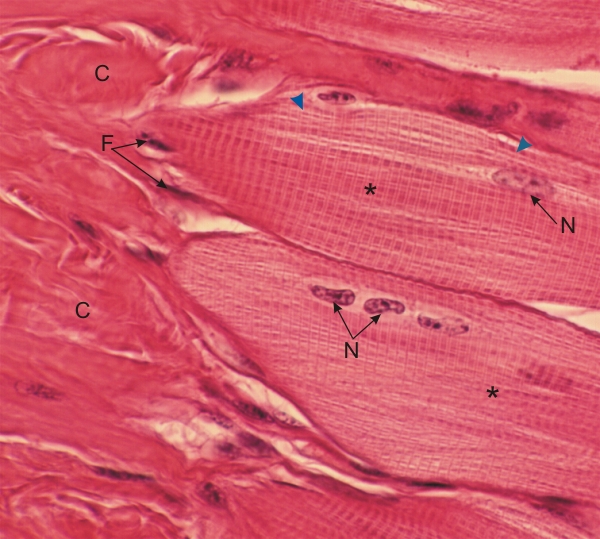
Section of the tongue showing a longitudinal section of two striated muscle fibres (*) inserted on a dense connective tissue.

The pointed conical extremities of the muscle cells (left) are closely attached to the dense type I collagen (C). In this section, the longitudinal views of the individual myofibrils are visible (arrowheads), together with their A- and I-bands, and the Z-lines in the I-bands.

The nuclei of muscle fibres (N) and of fibrocytes (F) are indicated.

Stain: H–­­E
Magnification: ×700


We'd very much appreciate receiving your comments and suggestions!We'd very much appreciate receiving your comments and suggestions!

Creative Commons Licence The text and images of this Histology Atlas, by Yves Clermont, Michael Lalli & Zsuzsanna Bencsath-Makkai, are licensed under a Creative Commons Attribution-Noncommercial-No Derivative Works 2.5 Canada Licence and cannot be modified without the written permission of the authors. Use of any text or images must carry an acknowledgement which includes a link to the original work.


HAweb/Zs B-M -> HAbase-generated on 2013-09-13