|
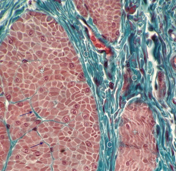
Section of the urinary bladder stained with Massons trichrome.
The bundle of muscle cells on the left (*) is seen in cross section while the small bundle on the lower right (+) is cut obliquely. The large bundles of smooth muscle fibres, some showing their nucleus, are separated by dense connective tissue septae (C) stained green.
The individual muscle fibres are separated by narrow spaces containing a few connective tissue fibres stained green (arrows).
Stain: Massons Trichrome
Magnification: ×900
|