Contents Yves Clermont, Michael Lalli, Zsuzsanna Bencsath-Makkai
LIGHT-MICROSCOPIC HISTOLOGY ATLAS
     3. Muscle Tissue
  1     2     3     4     5     6     7     8     9     10     11     12     13     14     15     16     17  

    3.4   Striated muscle Version française
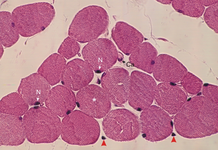
Cross section of a group of skeletal muscle fibres from the trachea.

In this muscle, the muscle cells have a circular profile. Their cytoplasm is packed with acidophilic myofibrils (*) seen in cross section. The nuclei (N) of the fibres are located at the periphery of their cytoplasms.

Between the muscle fibres is a small amount of connective tissue with a few fibrocytes (arrowheads). A capillary (Ca) is indicated.

Stain: H–­­E
Magnification: ×600


We'd very much appreciate receiving your comments and suggestions!We'd very much appreciate receiving your comments and suggestions!

Creative Commons Licence The text and images of this Histology Atlas, by Yves Clermont, Michael Lalli & Zsuzsanna Bencsath-Makkai, are licensed under a Creative Commons Attribution-Noncommercial-No Derivative Works 2.5 Canada Licence and cannot be modified without the written permission of the authors. Use of any text or images must carry an acknowledgement which includes a link to the original work.


HAweb/Zs B-M -> HAbase-generated on 2013-09-13