|
||
| 3. Muscle Tissue | ||
| 1 2 3 4 5 6 7 8 9 10 11 12 13 14 15 16 17 |
| |||
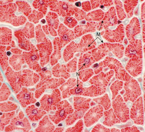 |
Section of a cardiac muscle showing transverse sections of cardiocytes.
These cardiac muscle fibres have irregular outlines (see Figure 3.1B). They show central nuclei (N). In the abundant cytoplasm the myofibrils are variable in dimension, the larger myofibrils being at the periphery of the cytoplasm (M). Cardiocytes are separated by a small amount of loose connective tissue (greenish). Stain: Massons Trichrome
|
||