|
||
| 3. Muscle Tissue | ||
| 1 2 3 4 5 6 7 8 9 10 11 12 13 14 15 16 17 |
| |||
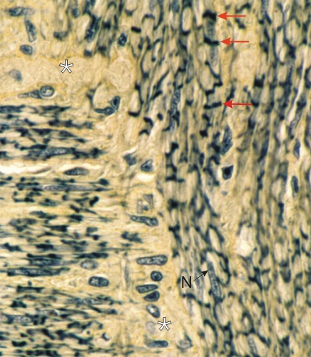 |
Section of the deferent duct (vas deferens) of the male reproductive system stained with TPA.
This field shows bundles of longitudinally sectioned smooth muscle fibres. In the bundles on the left, the muscle fibres are arranged horizontally. In the bundles on the right, the muscle fibres are arranged vertically. The smooth muscle fibres of the bundles on the right are contracted and show irregular, wavy, TPA-positive transverse bands (arrows). These contraction bands are not identical to the striations of skeletal muscle fibres. They appear to be due to the alignment, during contraction, of the dense cytoplasmic bodies on which the myofilaments are inserted (see Figure 3.1C). The nucleus (N) of a smooth muscle fibre and the yellow stained connective tissue (*) are identified. Stain: TPA
|
||