|
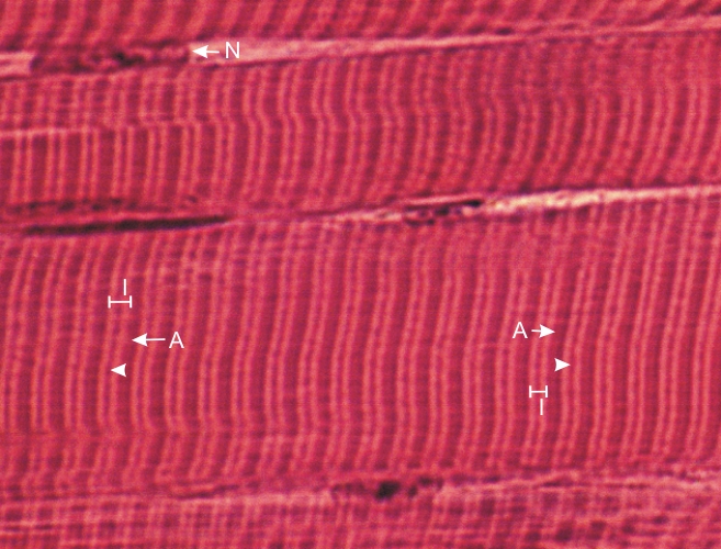
Longitudinal sections of two adjacent skeletal muscle fibres.
The A-bands (A) and the I-bands (I) are labelled. In the middle of the I-bands, the thin Z-lines are clearly seen (arrowheads). Note that, owing to the thickness of the section, the individual myofibrils are not distinct. Nevertheless, the A-bands and I-bands of adjacent myofibrils are clearly aligned across the width of the muscle fibres.
One nucleus (N) is labelled at the periphery of the muscle fibre above.
Stain: HE
Magnification: ×1000
|