Contents Yves Clermont, Michael Lalli, Zsuzsanna Bencsath-Makkai
LIGHT-MICROSCOPIC HISTOLOGY ATLAS
     3. Muscle Tissue
  1     2     3     4     5     6     7     8     9     10     11     12     13     14     15     16     17  

    3.2   Striated muscle Version française
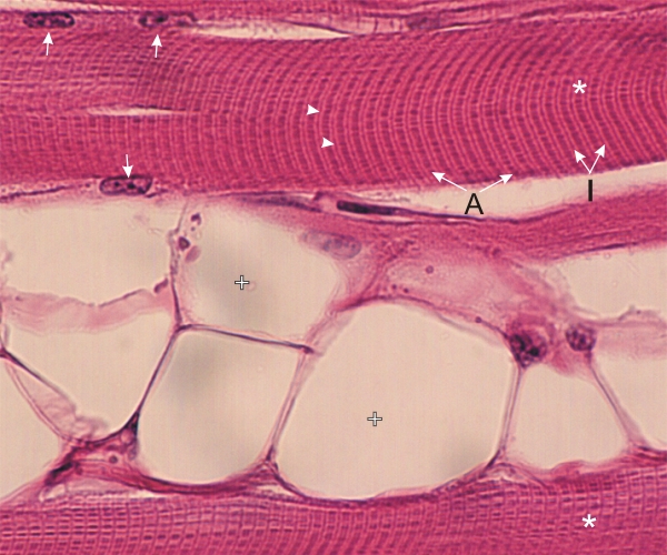
Longitudinal section of two skeletal muscle fibres (*) separated by adipocytes (+).

The muscle fibre (top) shows the characteristic cross striations of the myofibrils with their dark bands (A) and light bands (I). In the middle of the latter, the thin Z-lines (arrowheads) are visible. Several nuclei (arrows) of the muscle cells are seen at the periphery of their cytoplasm.

Stain: H–­­E
Magnification: ×600


We'd very much appreciate receiving your comments and suggestions!We'd very much appreciate receiving your comments and suggestions!

Creative Commons Licence The text and images of this Histology Atlas, by Yves Clermont, Michael Lalli & Zsuzsanna Bencsath-Makkai, are licensed under a Creative Commons Attribution-Noncommercial-No Derivative Works 2.5 Canada Licence and cannot be modified without the written permission of the authors. Use of any text or images must carry an acknowledgement which includes a link to the original work.


HAweb/Zs B-M -> HAbase-generated on 2013-09-13