Contents Yves Clermont, Michael Lalli, Zsuzsanna Bencsath-Makkai
LIGHT-MICROSCOPIC HISTOLOGY ATLAS
     3. Muscle Tissue
  1     2     3     4     5     6     7     8     9     10     11     12     13     14     15     16     17  

    3.11   Cardiac muscle Version française
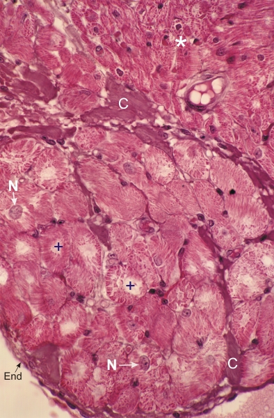
Section of a human cardiac muscle next to its endocardial surface (End).

This field shows, in cross or oblique sections, a collection of large cardiocytes (+). Standard-sized cardiocytes are seen at the top of the field (*). The large cardiocytes below have a large pale central cytoplasm, a few showing a nucleus (N). The acidophilic myofibrils occupy the periphery of the cells.

These large cardiocytes belong to an atrioventricular bundle considered to be part of the conducting system of the heart controlling the sequential rhythmicity of the atria and ventricles.

Some intensely stained dense connective tissue (C) is also indicated

Stain: H–­­E
Magnification: ×500


We'd very much appreciate receiving your comments and suggestions!We'd very much appreciate receiving your comments and suggestions!

Creative Commons Licence The text and images of this Histology Atlas, by Yves Clermont, Michael Lalli & Zsuzsanna Bencsath-Makkai, are licensed under a Creative Commons Attribution-Noncommercial-No Derivative Works 2.5 Canada Licence and cannot be modified without the written permission of the authors. Use of any text or images must carry an acknowledgement which includes a link to the original work.


HAweb/Zs B-M -> HAbase-generated on 2013-09-13