 |
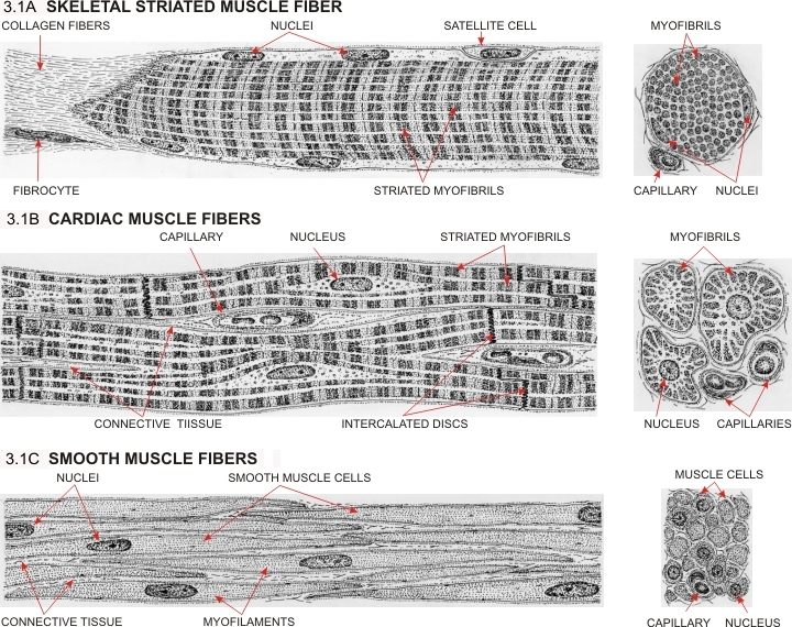
Figure 3.1A. Skeletal muscle fibres are large and long multinucleated cells. Their nuclei are located at the periphery of the cytoplasm. Their plasma membrane is covered by a basement membrane, inside of which are occasional satellite cells. Satellite cells serve to regenerate damaged muscle fibres. The cytoplasm is occupied by numerous cylindrical myofibrils showing the A- and I-bands (see details in Figure 3.6) forming the cross-striations. At the extreme left, the conical extremity of the muscle fibre shows numerous fine processes attached to the collagen fibres of a tendon or of a septum.
Figure 3.1B. Drawing of a few cardiac muscle fibres or cardiocytes. These elongated cells are relatively short and pleomorphic. They are attached tip to tip at the level of the intercalated discs. One or two nuclei are centrally located in each cardiocyte. The myofibrils are sometimes large and show irregular cross-sectional profiles. The cardiac muscle fibres are covered by a thin basement membrane which is absent, however, from the intercalated discs.
Figure 3.1C. Drawing showing a fascicle of fusiform smooth muscle fibres. These cells have only one nucleus and their cytoplasm contains myofilaments which are not grouped into striated myofibrils. These myofilaments are inserted onto miniscule fusiform bodies distributed throughout the cytoplasm or as plaques attached to the internal surface of the plasma membrane. Such cytoplasmic densities can be seen clearly only with an electron microscope. The surfaces of the cells are associated with a thin basement membrane.
|