|
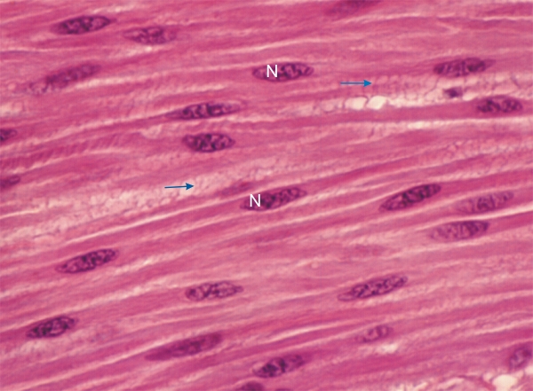
Section of the small intestine of a dog showing a bundle of smooth muscle fibres cut longitudinally.
Each cell has a long spindle-shaped cytoplasm with a single centrally located nucleus (N). The cytoplasm is filled with acidophilic non-striated contractile filaments oriented in the long axis of the cells. These filaments are not grouped into myofibrils.
The smooth muscle fibres are separated by reticular fibres (arrows) composed of type III collagen (see Figure 3.15.).
Stain: HE
Magnification: ×900
|