Sommaire Yves Clermont, Michael Lalli, Zsuzsanna Bencsath-Makkai
ATLAS D'HISTOLOGIE EN MICROSCOPIE OPTIQUE
     15. Appareil Reproducteur Mâle
  1     2     3     4     5     6     7     8     9     10     11     12     13     14     15     16     17     18     19     20     21     22     23     24     25  
  26     27     28     29     30     31     32     33     34     35     36     37     38     39     40     41     42     43     44  
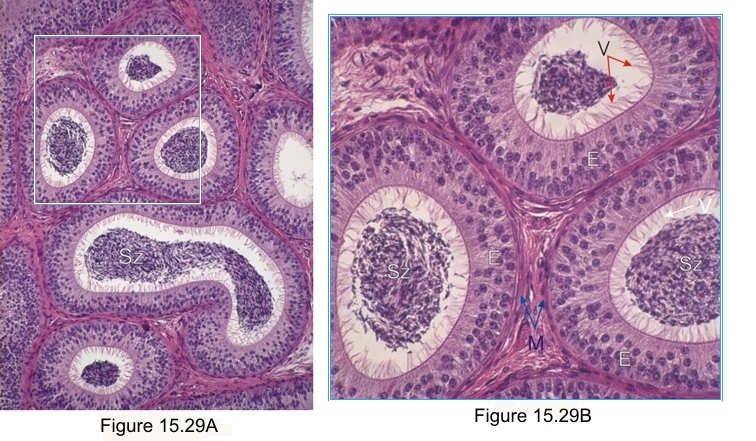
    15.29   Epididyme English version
Coupes du corps de l’épididyme d’un singe. Chez cette espèce l’épithélium, posé sur les cellules myoïdes de la membrane limitante(M), montre des cellules épithéliales prismatiques d’ une hauteur identique et régulière mais dont les noyaux occupent des niveaux différents dans leurs cytoplasmes. À leurs sommets de longs faisceaux de villosités (V) ou stéréocils qui rejoignent les spermatozoïdes (Sz).

Coloration: H–­­É
Grossissement: ×300; ×900


We'd very much appreciate receiving your comments and suggestions!We'd very much appreciate receiving your comments and suggestions!

Creative Commons Licence The text and images of this Histology Atlas, by Yves Clermont, Michael Lalli & Zsuzsanna Bencsath-Makkai, are licensed under a Creative Commons Attribution-Noncommercial-No Derivative Works 2.5 Canada Licence and cannot be modified without the written permission of the authors. Use of any text or images must carry an acknowledgement which includes a link to the original work.


HAweb/Zs B-M -> HAbase-generated on 2013-09-13