Sommaire Yves Clermont, Michael Lalli, Zsuzsanna Bencsath-Makkai
ATLAS D'HISTOLOGIE EN MICROSCOPIE OPTIQUE
     15. Appareil Reproducteur Mâle
  1     2     3     4     5     6     7     8     9     10     11     12     13     14     15     16     17     18     19     20     21     22     23     24     25  
  26     27     28     29     30     31     32     33     34     35     36     37     38     39     40     41     42     43     44  
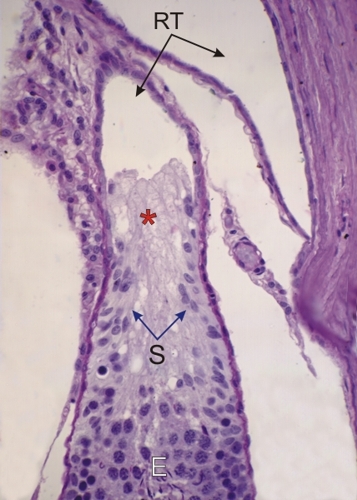
    15.20   Le rete testis English version
Coupe du testicule d’un rat montrant la jonction d’un tube séminifère avec le rete testis (RT).

Le rete testis est un réseau de canaux anastomosés bordé par un épithélium simple cubique.

À la jonction du tube séminifère au rete testis, l’épithélium séminifère (E) est remplacé par un épithélium composé exclusivement de cellules de Sertoli (S). Celles-ci forment une papille (*) qui occupe la lumière du rete testis sur une courte distance.

Coloration: H–­­É
Grossissement: ×400


We'd very much appreciate receiving your comments and suggestions!We'd very much appreciate receiving your comments and suggestions!

Creative Commons Licence The text and images of this Histology Atlas, by Yves Clermont, Michael Lalli & Zsuzsanna Bencsath-Makkai, are licensed under a Creative Commons Attribution-Noncommercial-No Derivative Works 2.5 Canada Licence and cannot be modified without the written permission of the authors. Use of any text or images must carry an acknowledgement which includes a link to the original work.


HAweb/Zs B-M -> HAbase-generated on 2013-09-13