Sommaire Yves Clermont, Michael Lalli, Zsuzsanna Bencsath-Makkai
ATLAS D'HISTOLOGIE EN MICROSCOPIE OPTIQUE
     15. Appareil Reproducteur Mâle
  1     2     3     4     5     6     7     8     9     10     11     12     13     14     15     16     17     18     19     20     21     22     23     24     25  
  26     27     28     29     30     31     32     33     34     35     36     37     38     39     40     41     42     43     44  
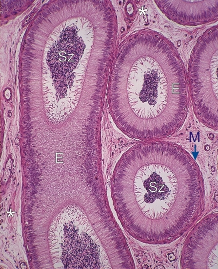
    15.26   Épididyme English version
Coupe de la tête de l’épididyme d’un singe.

Le canal épididymaire, très contourné, est vu en coupe longitudinale (à gauche) et en coupes transversales (à droite).

La lumière du canal contient de nombreux spermatozoïdes (Sz).

L’épithélium (E) est composé de grandes cellules prismatiques de taille identiques. Cet épithélium est posé sur une membrane limitante épaisse (M) formée de cellules myoïdes contractiles.

Les espaces intertubulaires sont occupés par un tissu conjonctif lâche (*) bien vascularisé.

Coloration: H–­­É
Grossissement: ×400


We'd very much appreciate receiving your comments and suggestions!We'd very much appreciate receiving your comments and suggestions!

Creative Commons Licence The text and images of this Histology Atlas, by Yves Clermont, Michael Lalli & Zsuzsanna Bencsath-Makkai, are licensed under a Creative Commons Attribution-Noncommercial-No Derivative Works 2.5 Canada Licence and cannot be modified without the written permission of the authors. Use of any text or images must carry an acknowledgement which includes a link to the original work.


HAweb/Zs B-M -> HAbase-generated on 2013-09-13