Sommaire Yves Clermont, Michael Lalli, Zsuzsanna Bencsath-Makkai
ATLAS D'HISTOLOGIE EN MICROSCOPIE OPTIQUE
     15. Appareil Reproducteur Mâle
  1     2     3     4     5     6     7     8     9     10     11     12     13     14     15     16     17     18     19     20     21     22     23     24     25  
  26     27     28     29     30     31     32     33     34     35     36     37     38     39     40     41     42     43     44  
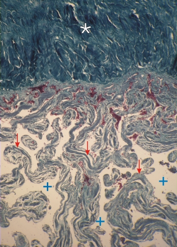
    15.44   Pénis humain English version
Coupe d’un pénis humain.

Cette coupe montre en haut du champ, intensément coloré en vert, le tissu conjonctif dense de l’albuginée (*) (tunica albuginea) du pénis.

Les espaces vasculaires (+) du corps caverneux (corpus cavernosum, voir la figure 15.1) sont separés par des trabécules ou lames de tissu conjonctif (flèches) contenant des faisceaux de cellules musculaires lisses.

Coloration: Trichrome de Masson
Grossissement: ×100


We'd very much appreciate receiving your comments and suggestions!We'd very much appreciate receiving your comments and suggestions!

Creative Commons Licence The text and images of this Histology Atlas, by Yves Clermont, Michael Lalli & Zsuzsanna Bencsath-Makkai, are licensed under a Creative Commons Attribution-Noncommercial-No Derivative Works 2.5 Canada Licence and cannot be modified without the written permission of the authors. Use of any text or images must carry an acknowledgement which includes a link to the original work.


HAweb/Zs B-M -> HAbase-generated on 2013-09-13