Sommaire Yves Clermont, Michael Lalli, Zsuzsanna Bencsath-Makkai
ATLAS D'HISTOLOGIE EN MICROSCOPIE OPTIQUE
     15. Appareil Reproducteur Mâle
  1     2     3     4     5     6     7     8     9     10     11     12     13     14     15     16     17     18     19     20     21     22     23     24     25  
  26     27     28     29     30     31     32     33     34     35     36     37     38     39     40     41     42     43     44  

    15.18   Cellules de Leydig English version
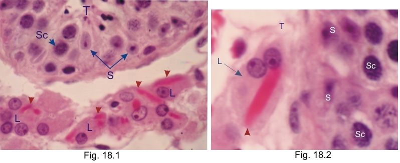
Cellules de Leydig (L) humaines montrant, dans leur cytoplasme, des éléments très acidophiles correspondants aux crystalloïdes (cristaux protéiques) de Reinke (pointes de flèches).

Les structures suivantes sont étiquetées: Les tubes séminifères (T), les noyaux des cellules de Sertoli (S) et les noyaux des spermatocytes (Sc).

Coloration: H–­­É
Grossissement: ×1000, ×1200


We'd very much appreciate receiving your comments and suggestions!We'd very much appreciate receiving your comments and suggestions!

Creative Commons Licence The text and images of this Histology Atlas, by Yves Clermont, Michael Lalli & Zsuzsanna Bencsath-Makkai, are licensed under a Creative Commons Attribution-Noncommercial-No Derivative Works 2.5 Canada Licence and cannot be modified without the written permission of the authors. Use of any text or images must carry an acknowledgement which includes a link to the original work.


HAweb/Zs B-M -> HAbase-generated on 2013-09-13