Sommaire Yves Clermont, Michael Lalli, Zsuzsanna Bencsath-Makkai
ATLAS D'HISTOLOGIE EN MICROSCOPIE OPTIQUE
     15. Appareil Reproducteur Mâle
  1     2     3     4     5     6     7     8     9     10     11     12     13     14     15     16     17     18     19     20     21     22     23     24     25  
  26     27     28     29     30     31     32     33     34     35     36     37     38     39     40     41     42     43     44  

    15.28   Epididyme de l’homme English version
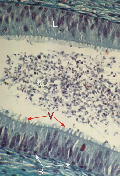
Coupe du canal épididymaire humain.

De nombreux spermatozoïdes (Sz) occupent la lumière du canal.

L’épithélium (E) est composé de hautes cellules prismatiques dont les noyaux sont à divers niveaux de leurs cytoplasmes. On observe à leur sommet des faisceaux de microvillosités ou stéréocils (V).

De nombreuses petites cellules épithéliales basales (B) sont associées à la membrane limitante du canal.

Coloration: Trichrome de Masson
Grossissement: ×800


We'd very much appreciate receiving your comments and suggestions!We'd very much appreciate receiving your comments and suggestions!

Creative Commons Licence The text and images of this Histology Atlas, by Yves Clermont, Michael Lalli & Zsuzsanna Bencsath-Makkai, are licensed under a Creative Commons Attribution-Noncommercial-No Derivative Works 2.5 Canada Licence and cannot be modified without the written permission of the authors. Use of any text or images must carry an acknowledgement which includes a link to the original work.


HAweb/Zs B-M -> HAbase-generated on 2013-09-13