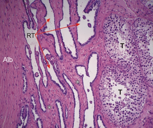
Sommaire Yves Clermont, Michael Lalli, Zsuzsanna Bencsath-Makkai
ATLAS D'HISTOLOGIE EN MICROSCOPIE OPTIQUE
     15. Appareil Reproducteur Mâle
  1     2     3     4     5     6     7     8     9     10     11     12     13     14     15     16     17     18     19     20     21     22     23     24     25  
  26     27     28     29     30     31     32     33     34     35     36     37     38     39     40     41     42     43     44  

    15.21   Rete testis English version
Coupe d’un testicule humain dans la région du hile.

(À droite) il y a quelques tubes séminifères (T) et (à gauche) l’albuginée ou tunica albuginea (Alb).

Entre l’albuginée et les tubes séminifères des canaux forment le rete testis (RT). Ces espaces irréguliers sont interconnectés les uns aux autres. Ils sont bordés d’un épithélium simple composé de cellules épithéliales de tailles variables qui sont étroitement associées au tissu conjonctif dense (*) qui sépare les canaux du rete testis.

Coloration: H–­­É
Grossissement: ×250


We'd very much appreciate receiving your comments and suggestions!We'd very much appreciate receiving your comments and suggestions!

Creative Commons Licence The text and images of this Histology Atlas, by Yves Clermont, Michael Lalli & Zsuzsanna Bencsath-Makkai, are licensed under a Creative Commons Attribution-Noncommercial-No Derivative Works 2.5 Canada Licence and cannot be modified without the written permission of the authors. Use of any text or images must carry an acknowledgement which includes a link to the original work.


HAweb/Zs B-M -> HAbase-generated on 2013-09-13