|
||
| 14. Glandes Endocrines | ||
| 1 2 3 4 5 6 7 8 9 10 11 12 13 14 15 16 17 18 19 20 21 22 23 24 25 | ||
| 26 27 28 29 30 |
| |||
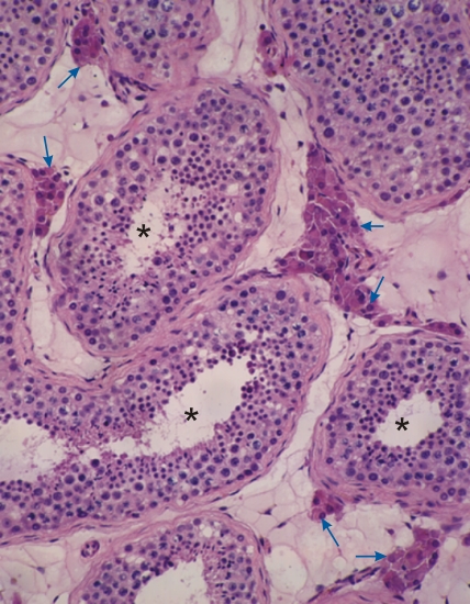 |
Coupe dun testicule humain. Dans le tissu conjonctif entre les tubes séminifères (*) on peut facilement identifier des groupes de cellules de Leydig, très chromophiles (flèches). Ces cellules endocrines sécrètent la testostérone. Coloration: HÉ
|
||