Sommaire Yves Clermont, Michael Lalli, Zsuzsanna Bencsath-Makkai
ATLAS D'HISTOLOGIE EN MICROSCOPIE OPTIQUE
     14. Glandes Endocrines
  1     2     3     4     5     6     7     8     9     10     11     12     13     14     15     16     17     18     19     20     21     22     23     24     25  
  26     27     28     29     30  

    14.19   Cortex d’une surrénale humaine English version
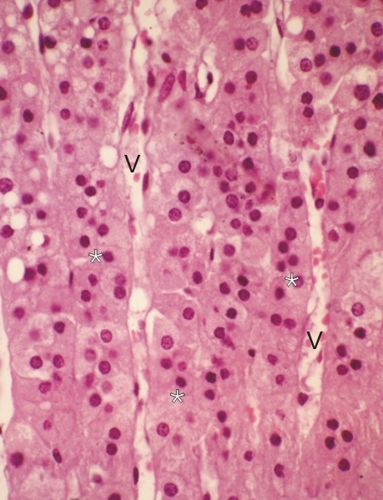
Ce champ montre la zone fasciculée du cortex d’une surrénale humaine.

Les cellules glandulaires, en rangées doubles (*), forment des cordons ou lames anastomosées orientés radialement dans le cortex.

Ces cordons ou lames sont séparés par des espaces conjonctifs contenant des capillaires ou sinus veineux (V).

Coloration: H–­­É
Grossissement: ×600


We'd very much appreciate receiving your comments and suggestions!We'd very much appreciate receiving your comments and suggestions!

Creative Commons Licence The text and images of this Histology Atlas, by Yves Clermont, Michael Lalli & Zsuzsanna Bencsath-Makkai, are licensed under a Creative Commons Attribution-Noncommercial-No Derivative Works 2.5 Canada Licence and cannot be modified without the written permission of the authors. Use of any text or images must carry an acknowledgement which includes a link to the original work.


HAweb/Zs B-M -> HAbase-generated on 2013-09-13